

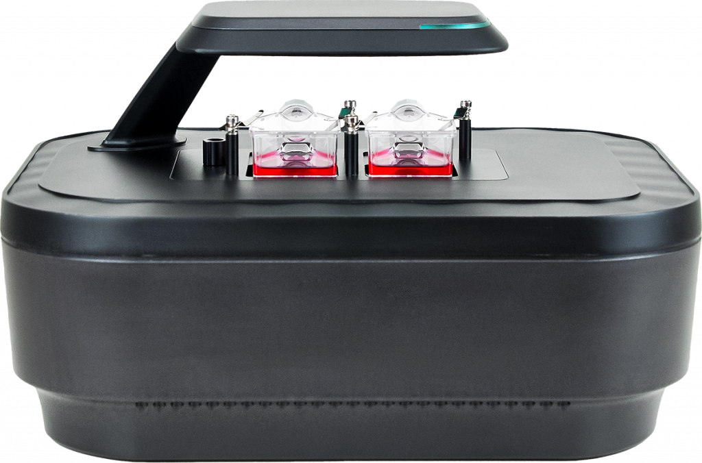
REAL-TIME CELL IMAGING SYSTEM
Fluorescent TimeLapse Device
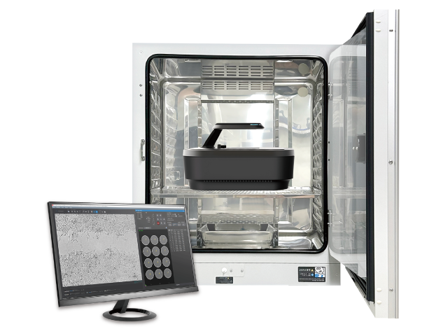
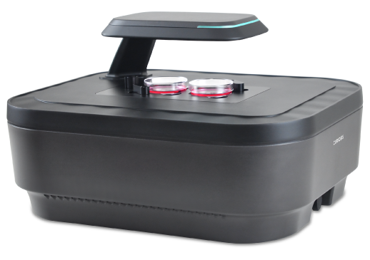
Automated Cell Imaging System Directly in Incubators
An innovative cellular imaging system designed to be placed directly within your CO2 incubator. With exceptional image quality and unparalleled ease of use, it provides researchers with advanced features. Options such as stitching, z-stacking, and the ability to focus differently based on positions offer users great flexibility in acquiring their images.
By enabling real-time cell monitoring inside the incubator, it allows for observation and tracking of cellular dynamics. The system’s dual-fluorescence and bright-field microscopy enable simultaneous visualization of multiple markers. Multipoint time-lapse imaging captures dynamic cellular events at different locations and scheduled times.
Image analysis is then automated through the software. It allows for the extraction of photos and videos, as well as proliferation, cytotoxicity, healing (scratch test) curves, fluorescence rates, and sizes and numbers of cells or 3D cellular structures. We custom-develop software modules to bring you closer to the cellular functions that interest you.
KEY
FEATURES


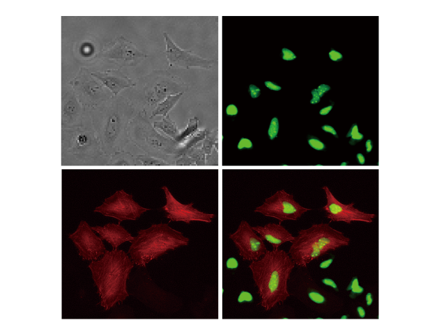
Multicolor fluorescence and brightfield imaging
- With its capabilities in multicolor fluorescence imaging and bright-field microscopy, our solutions enable the capture of high-quality and high-resolution images.
- With enhanced scanning methods and innovative merging techniques, the system reduces scanning time, enabling researchers to analyze cellular dynamics with exceptional clarity and efficiency.
Real-time monitoring inside the incubator
- Designed to facilitate real-time monitoring of cells inside an incubator, by simply placing the device inside the incubator and connecting it to an external PC, researchers can remotely observe cells in real-time.
- With the time-lapse function, cell images are captured according to the schedule set by the researcher; the images can then be easily converted into time-lapse videos.

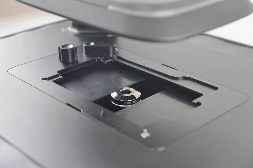
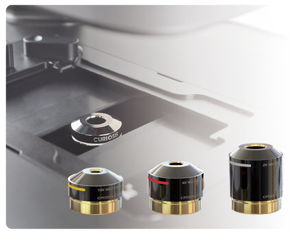
User-interchangeable objective lens
- User-interchangeable objectives provide flexibility to researchers based on their specific study needs. With options such as 2X, 4X, and 10X objectives, users can manually switch between objectives as needed.
Capturing images from multiple positions
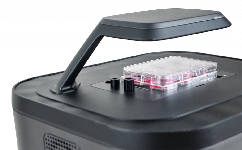
- Enabling imaging of samples from multiple positions by automatically moving the integrated camera beneath the fixed culture support. This ensures a stable environment for cells, resulting in improved image quality and precise research outcomes.
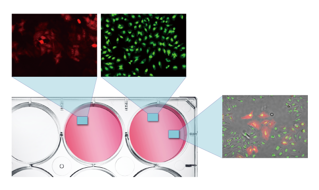
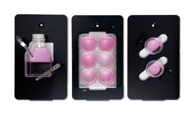
Compatibility with all culture supports (all brands).
- The system is compatible with various cell culture supports such as multi-well plates (up to 96 wells), flasks, Petri dishes, and slides, and can switch between them by simply replacing the adapters as needed.
Specification
| Imaging modes | Brightfield, Dual fluorescence (Green & Red) |
|---|---|
| Objective Lens | 2X, 4X, 10X (User-interchangeable) |
| Fluorescence | Green : EX (470/40), EM (540/50) Red : EX (562/40), EM (641/75) |
| Stage | Fully motorized XYZ (Fixed stage, camera moving type) |
| Camera | High sensitivity 5.0 MP CMOS |
| Imaging positions | Multiple |
| Field-of-view | 2X (2.08 x 1.55 mm), 4X (1.46 x 1.09 mm), 10X (0.72 x 0.54 mm) |
| Focus | Autofocus, Manual focus |
| Imaging methods | Single/multicolor, stitching, stacking, time-lapse, real-time recording |
| Included software | Scan App, Analysis App |
| Dimensions (H x W x L) | 250 x 338 x 412 mm |
| Weight | 9.6 kg |
| Culture vessels | Well plate up to 96-well, flask, dish, slide |
| File export format | TIFF, AVI (JPEG, PNG) |
| Operating environment | 10~40℃, 20~95% humidity |
| Power requirement | 100-240V, ~50/60Hz |
| O/S required | Windows 10 and above |
| Incubator specification | Above 200L (recommended) |
Ordering information
| Catalog No. | Description |
|---|---|
| CRCLG-P01 | Celloger® Pro, Live cell imaging system(Bright Field, GFP+RFP) & Objective Lens set |
| CRCLG-PL02A | Objective lens (2X) |
| CRCLG-PL04A | Objective lens (4X) |
| CRCLG-PL10A | Objective lens (10X) |
| CRCLG-PLS | Objective lens set (2X, 4X, 10X) |
| CRCLG-MPWPS | Vessel holder, Well plate 6~96 (Single) |
| CCRCLG-MPTFS25 | Vessel holder, T-FlaskA25cm2 (Single) |
| CRCLG-MPTFD25 | Vessel holder, T-FlaskA25cm2 (Dual) |
| CRCLG-MPTFS75 | Vessel holder, T-FlaskA75cm2 (Single) |
| CRCLG-MPPDD35 | Vessel holder, Petri dish 35mm (Dual) |
| CRCLG-MPPDD60 | Vessel holder, Petri dish 60mm (Dual) |
| CRCLG-MPPDS90 | Vessel holder, Petri dish 90/100mm (Single) |
| CRCLG-MPSH03 | Vessel holder, Slide (Triple) |


