
3D cell imaging by tomography

Estier is a high-level optical coherence tomography (OCT) instrument that allows non-invasive analysis of 3D structures, including microvessels/tubular structures, tissues, spheroids and organoids.
As OCT provides an advantageous platform to conveniently visualise the in vitro and ex vivo dynamics of 3D structures, it can be used as an alternative visualisation/monitoring method to obtain quantitative information on morphological changes occurring in 3D cell culture samples.
Estier OCT technology enables label-free, non-invasive tomographic imaging and monitoring of microvessels, neovascularisation formation to quantify and assess morphological changes in a short time. Ideal for tumour invasion testing and molecule efficacy assessment.
The ability to measure the actual volume (X, Y and Z directions) of 3D structures (spheroids/organoids) allows true comparison of drug efficacy in preclinical models.
Detects necrotic regions and quantifies volume, internal cavities, tubular structures, etc., with impressive focus.
Specifications
| Data Output parameters | Tomogram in user indicated location / 3D image from user indicated view point / Movie output of tomogram / Animation output of 3D image / Quantified value: distance between point to point, area of 2D image, volume, sphericity, surface rough degree, cavity volume |
| Resolution | High resolution: 3 μm, Low resolution: 10 μm |
| Max. FOV | High resolution: 1 x 1 mm, Low resolution: 10 x 10 mm (Wide F.O.V.) |
| Max. depth | High resolution / Low resolution: 1,000 μm (according to sample) |
| Observation time | Cross-sectional observation: 0.5 sec. or more 3D observation: High resolution 0.3 x 0.3 x 0.3 mm / 3 µm: 1 min. Low resolution 5.0 x 5.0 x 1.0 mm / 10 µm: 9 min. |
| Vessel | Micro well plate, Culture dish, etc. |
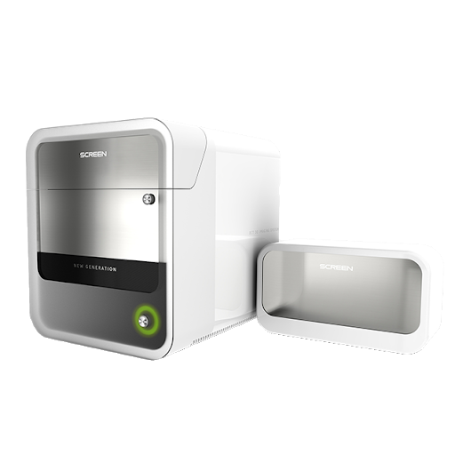
| Components | Main unit (W20 x D20 x H19 inch) Sub unit (W7 x D18 x H12 inch) PC (W7 x D18 x H17 inch) + mouse, key board, joy-stick |
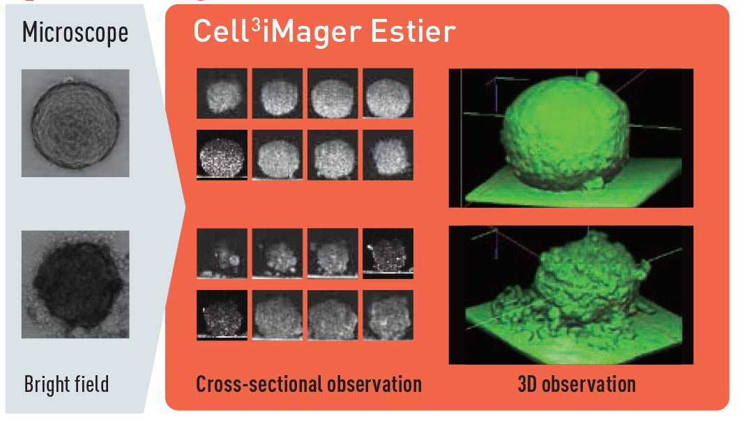
- 3D morphological analysis of cultures / internal structures with OCT. OCT is a non-invasive technology that can be used to acquire the tomogram of a living cell or organism using weak near infrared light.
- Time-lapse observation by non-invasive and label-free analysis Real-time observation of spheroid formation at collapse after addition of an anti-cancer drug Detailed observation of the 3D structure of spheroids that conventional microscopes cannot recognise
- Compatible with all standard micro well plates (SBS format) and culture dishes Specific well plates optimised for 3D culture, including Corning® Elplasia®, can be used for imaging SCREEN offers customised solutions to our researchers. These include the implementation of custom plate formats, such as microfluidic chips, organ-on-chips.