

REAL-TIME CELL IMAGING SYSTEM
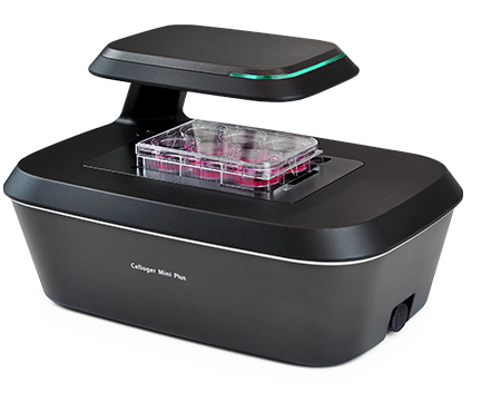
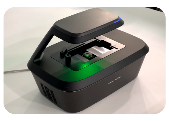
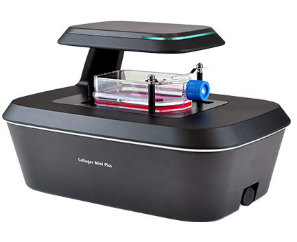
Timelapse Mini+
Empower your research…
Real-time quantitative time-lapse cell imaging system, based on brightfield microscopy and also equipped with fluorescence.
Resistant to temperature and humidity, it can be placed directly inside conventional CO2 or hypoxia incubators.
Serial image acquisition allows researchers to observe cell morphology and dynamics in real time over long periods, directly inside the incubator. Stitching options, Z-stacking, and the ability to set different focus positions give users great flexibility in image acquisition.
Image analysis is then automated by the software.
It enables extraction of photos and videos, as well as proliferation, cytotoxicity, wound healing (scratch test) curves, fluorescence intensity, and the size and number of cells or 3D cellular structures. We develop customized software modules to match as closely as possible the cellular functions you are interested in.
KEY FEATURES


REAL-TIME MONITORING
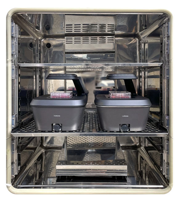
You can easily monitor live cells inside the incubator for long periods without disturbing the environment required for cell culture.
Compatible with all CO2 and hypoxia incubators.
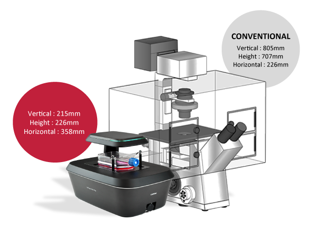
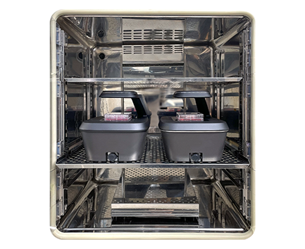
COMPACT SIZE
- Compact size: 226 (H) x 358 (L) x 215 (W) mm
- Multiple systems can fit inside a standard CO2 incubator.
- As the system weighs approximately 5 kg, it can be easily moved in and out of the incubator.
MULTI-POSITION IMAGING
- Using the motorized camera that moves 117 mm x 77 mm along the x and y axes respectively, multiple positions can be captured following the schedule (intervals, cycles, total time) defined by the researcher.
STABLE IMAGING PERFORMANCE
- No movement of the culture vessel (neither during imaging nor when retrieving the plate). The camera inside the system moves to capture cell images at multiple positions. The cell sample remains stable in an environment conducive to cell growth.

TIME-LAPSE VIDEO
- The images captured according to the defined program can be converted into videos in just a few simple clicks.
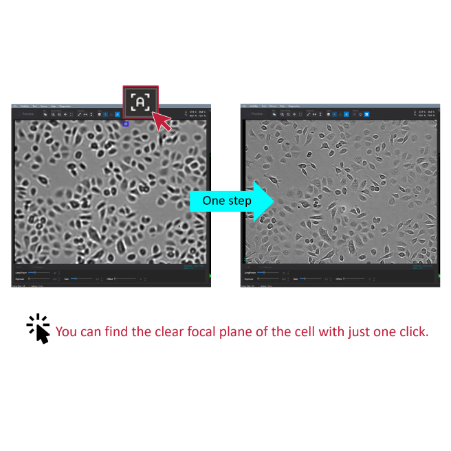
Autofocus
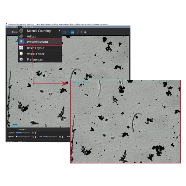
Recording preview
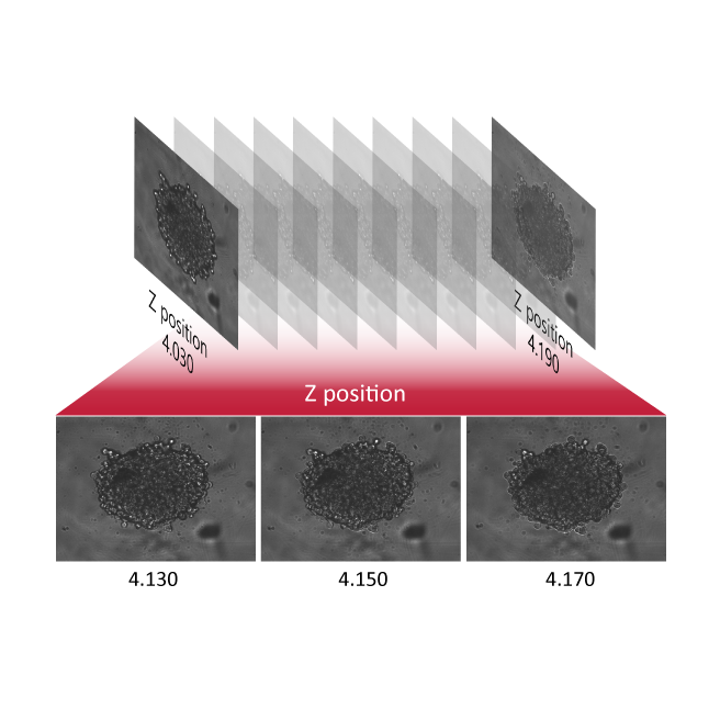
Z-stacking
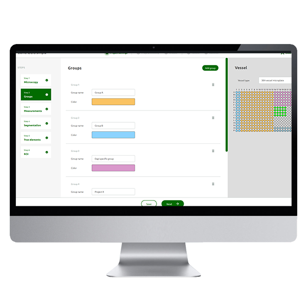
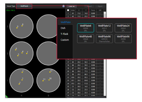
Software
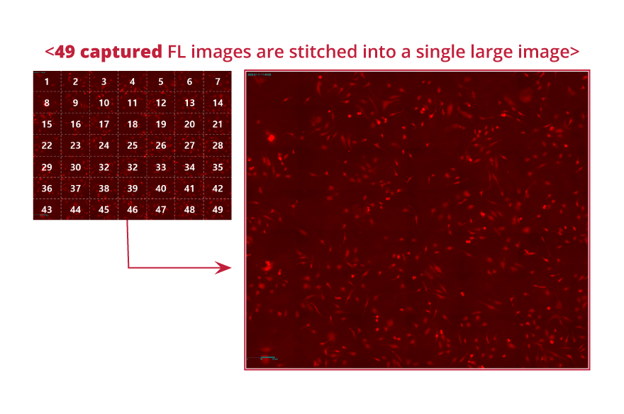
Image stitching
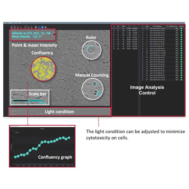
Analysis tools
Specifications
| Dimensions (H x W x L) | 226 x 215 x 358 mm |
|---|---|
| Weight | 5.6 kg / 12.3 lb |
| Objective lens | 4X / 10X |
| Imaging modes | Brightfield, Fluorescence (Green / Red) |
| Fluorescence | Green: Excitation (470/40x) / Emission (510lp) Red: Excitation (525/30x) / Emission (570lp) |
| Light source | LED |
| Camera | 5 MP CMOS |
| Stage | Motorized XYZ |
| Imaging positions | Multiple |
| File export format | TIFF, AVI (JPEG, PNG) |
| Culture vessels | Flask, dish, well plate, slide |
| Operating environment | 10–40°C, 20–95% humidity |
| Power requirements | 100–240V, ~50/60Hz |


